4 sierpnia, 2014
4 sierpnia, 2014
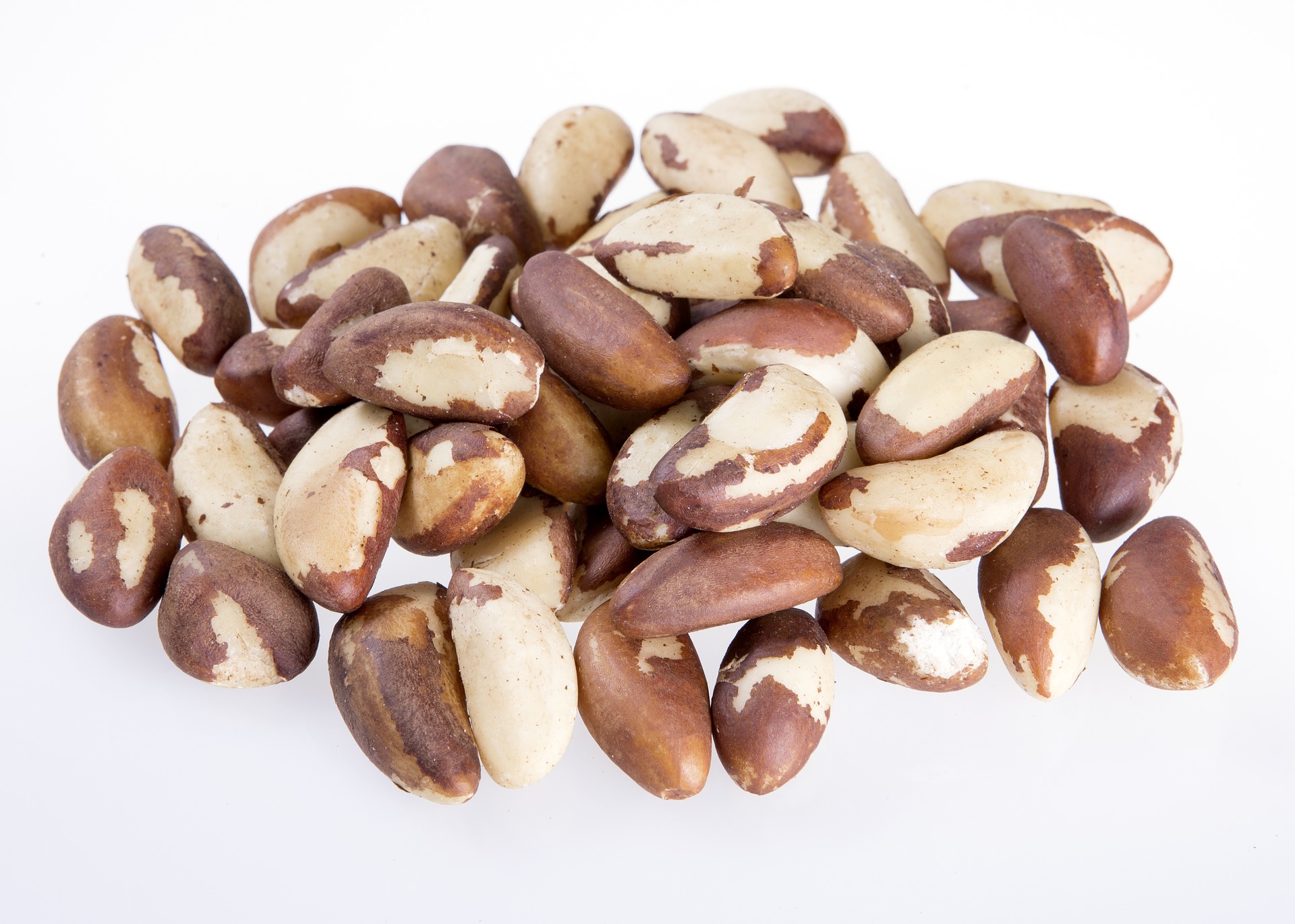
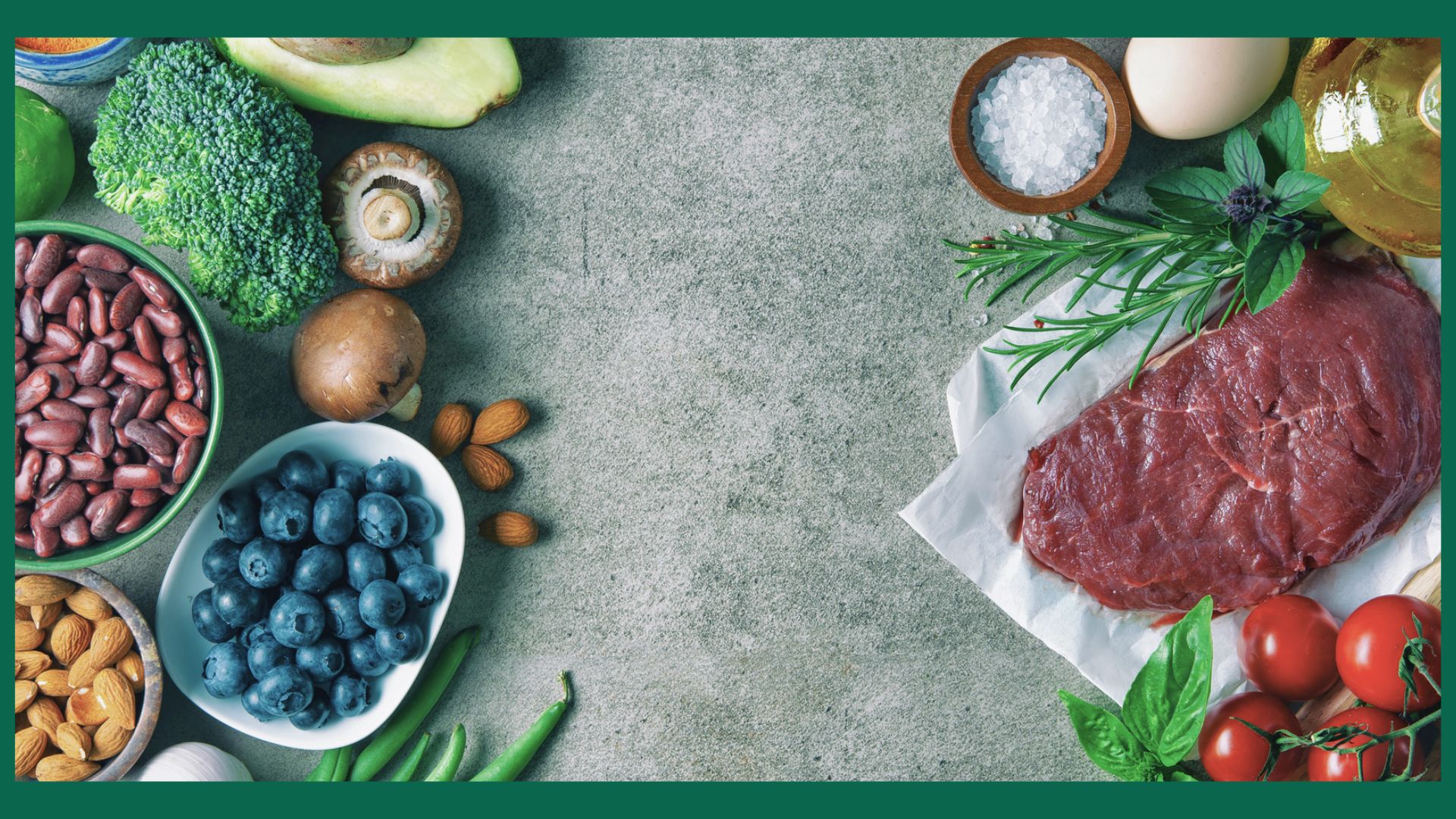
Selen jest niezbędnym śladowym minerałem dla poprawnego metabolizmu hormonów tarczycy, optymalnej odpowiedzi systemu odpornościowego, płodności, rozwoju mięśni i ochrony antyoksydacyjnej. Na początku XIX wieku gdy odkryto selen był uznany za toksyczną substancję. Dopiero w 1957 roku, Klaus Schwarz, biochemik Narodowych Instytutów Zdrowia, odkrył że szczury z niedoborami witaminy E są chronione uszkodzeniem wątroby przez selen. Rok później w 1958 roku, naukowcy z uniwersytetu w Oregon odkryli, że niedobory selenu są odpowiedzialne za degenerację mięśni u bydła pasącego się na terenach gdzie gleba jest uboga w selen. Suplementacja selenem pozwoliła zapobiec chorobie „białych mięśni”.
W latach 60 tych i 70 tych w Chinach przeprowadzane były badania nad chorobą Keshans, która doprowadza do uszkodzenia mięśnia sercowego. Również udowodniono, że suplementacja selen zapobiega rozwoju choroby. W latach 80 tych w USA rozpoznano niedobory selenu jako przyczyną śmierci w wyniku kardiomiopatii.
Selen jest faktycznie niezbędnym minerałem, ale to co nas interesuje to selenoproteiny. To one są zaangażowane w ochronę antynowotworową, antyoksydacyjną, płodność czy metabolizm hormonów tarczycy. Selenocysteina powstaje z zastąpienia cząsteczki siarki selenem. Selenocysteina w sekwencji aminokwasów daje selenoproteiny. Vadim N. Gladyshev zajmuje się korektą selenocysteiny w oryginalnej strukturze DNA i śledząc jego pracę można się wiele dowiedzieć na temat selenocysteiny.
Selenoproteiny wciąż nie są jednak dobrze zbadane, jednak parę znamy:
2) http://www.ncbi.nlm.nih.gov/pubmed/23046013
In patients with Hashimoto’s disease and in pregnant women with anti-TPO antibodies, selenium supplementation decreases anti-thyroid antibody levels and improves the ultrasound structure of the thyroid gland.
U pacjentów z Chorobą Hashimoto i ciężarnych kobiet mających pzreciwciała TPO, suplementacja selenem zmniejszyła poziom przeciwciała traczycowych i poprawiła strukturę tarczycy w obrazie USG.
3) http://www.ncbi.nlm.nih.gov/pubmed/20039895
Our study aimed to investigate whether physiological doses of selenium (Se) influence the natural course of autoimmune thyroiditis (AIT). DESIGN AND PATIENTS: A total of 76 consecutive patients (65 F, 11 M, median 43, range 15-75 years) with AIT, normal or slightly elevated TSH and fT4 within the normal range were divided into two groups:
Group 0 (30 cases) was given no treatment while
Group 1 (46 cases) was treated with sodium selenite 80 μg/day as a single oral dose for 12 months.
Thyroperoxidase and thyroglobulin autoantibodies (TPO-Ab; Tg-Ab), TSH, fT4 and urine iodine concentrations (UIC) were measured at baseline and after 6 and 12 months of follow-up. Thyroid ultrasonography (US) was performed at each follow-up point. Echogenicity was measured by histographic analysis of gray-scale pixels (gsp) ranging from 0 = black to 255 = white.
RESULTS:
Thyroid echogenicity decreased significantly in both groups after 6 months, but after 12 months, it had changed no more in Group 1, whereas it had dropped further in Group 0. No significant variation in TPO-Ab or Tg-Ab levels was observed between the two groups after 6 months, but both values decreased significantly after 12 months in Group 1, and five patients in this group became negative for TPO-Ab. TSH and FT4 showed no significant variations in either group.
CONCLUSIONS: Dietary supplementation with physiological doses of Se seems to be effective in preventing a reduction in thyroid echogenicity after 6 months of treatment and in reducing TPO-Ab and Tg-Ab after 12 months, but does not modify TSH or FT4.
Wnioski: Suplementacja seleny wydaje się być skuteczna w zapobieganiu i redukcji echogeniczności tarczycy po 6 mc leczenia i reduckji prziciwał TPO i TG po 12 miesiącach bez zmiany TSH i fT4.
4) http://www.ncbi.nlm.nih.gov/pubmed/23158484
To evaluate the effects of selenium (Se) supplementation on concentrations of thyroid peroxidase antibodies (TPOAb) and TPOAb IgG subclasses in autoimmune thyroiditis (AIT) patients with different thyroid functional status.
METHODS: A blind and placebo-controlled prospective study was performed for a total of 134 cases with AIT and thyroid peroxidase antibodies above 300 U/ml. Their mean age was 41 years (range: 15-70). All of them were recruited from Department of Endocrinology, First Affiliated Hospital of China Medical University from June 2008 to June 2009 and divided into
2 groups according to thyroid function:
euthyroidism or subclinical hypothyroidism (n = 89) and
hypothyroidism (n = 45).
Then they were randomized into 2 groups:
selenium-treated and placebo-treated.
And 49 cases in subclinical autoimmune thyroiditis group and 28 cases in hypothyroidism group received 200 µg oral selenium yeast daily for 6 months while others placebo. Serum concentrations of TPOAb, TPOAb IgG subclasses, thyroid-stimulating hormone (TSH), free thyroxine (FT(4)) and Se were measured at baseline and after 3 and 6 months of follow-up.
RESULTS: The TPOAb levels showed an overall decrease of 4.3% at 3 months and of 12.6% at 6 months (both P < 0.05) post-supplementation in subclinical autoimmune thyroiditis patients. In overt hypothyroidism patients, the overall decrease of TPOAb concentrations was 21.9% at 3 months and 20.4% at 6 months (both P < 0.05) compared with those at pre-treatment. The predominant TPOAb IgG subclasses in sera from the AIT patients were IgG1, IgG3 and IgG4 and the positive percentages 72%, 41% and 72% respectively. The positive rate and concentrations of IgG3 in the patients with hypothyroidism were significantly higher than those of subclinical autoimmune thyroiditis (P < 0.05). Significant decreases in IgG1 and IgG3 levels were noted in subclinical autoimmune thyroiditis group at 6 months post-supplementation (P < 0.05). IgG1 levels in overt hypothyroidism decreased significantly compared with those at pre-supplementation (P < 0.05). In all patients with supplementation (n = 77), the TPOAb levels decreased in 52 at 6 months while increase or no change occurred in 25. The positive percentage and concentrations of IgG1 in patients whose TPOAb levels decreased at 6 months post-supplementation were markedly higher than those whose TPOAb levels increased (P < 0.05).
CONCLUSION: Se is effective in reducing TPOAb concentrations and the predominant decreasing TPOAb IgG subclasses are IgG1 and IgG3. And a high level of IgG1 subclass may explain the difficult decline of TPOAb.
Wnioski: Selen jest efektywny w obniżeniu przecieciał TPO, w tym obniżeniu dominujących podklas IgG1 i IgG3.
5) http://www.ncbi.nlm.nih.gov/pubmed/17450242
Hashimoto’s thyroiditis and the role of selenium. Current concepts.Mazokopakis EE, Chatzipavlidou V.
Hashimoto’s thyroiditis (HT) is part of the spectrum of autoimmune thyroid diseases. Clinical manifestations of HT are variable and commonly include diffuse or nodular goiter with euthyroidism, subclinical hypothyroidism and permanent hypothyroidism. Uncommonly, HT causes acute destruction of thyroid tissue and release of stored thyroid hormones, causing transient thyrotoxicosis (hashitoxicosis). The contribution of methods and techniques of nuclear medicine to diagnosis and differential diagnosis of HT is indisputable. In HT patients with overt hypothyroidism L-thyroxine (L-T(4)) should be given in the usual replacement doses, but in HT patients with a large goiter and normal or elevated serum thyroid-stimulating hormone (TSH), L-T(4) may be given in doses sufficient to suppress serum TSH. Symptomatic patients with hashitoxicosis and low 24-hour thyroid radioactive iodine ((123)I or (123)I) uptake (RIU) may be treated with beta-blockers (as propranolol) and sodium ipodate or iopanoic acid (iodinated contrast agents) that block the peripheral conversion of T(4) to T(3). Recent clinical studies have documented the suppressive effect of selenium treatment on serum anti-thyroid peroxidase concentrations in patients with HT.
Wnisoki: Badania pokazują hamujący efekt suplementacji selenem na poziom anty TPO u pacjentów chorych na Hashimoto.
6) http://www.ncbi.nlm.nih.gov/pubmed/21508145
No previous study determined monocyte- and lymphocyte-suppressing effects of levothyroxine and selenomethionine and assessed whether their coadministration is superior to treatment with only one of these drugs.
OBJECTIVE:
Our objective was to compare the effect of levothyroxine and selenomethionine on monocyte and lymphocyte cytokine release and systemic inflammation in patients with Hashimoto’s thyroiditis.
DESIGN, SETTING, PARTICIPANTS, AND INTERVENTION:
We conducted a randomized clinical trial involving a group of 170 ambulatory euthyroid women with recently diagnosed and previously untreated Hashimoto’s thyroiditis and 41 matched healthy subjects. Participants were randomized in a double-blind fashion to receive a 6-month treatment with levothyroxine, selenomethionine, levothyroxine plus selenomethionine, or placebo. One hundred sixty-five patients completed the study.
MAIN OUTCOME MEASURES:
Monocyte and lymphocyte release of proinflammatory cytokines and plasma levels of C-reactive protein (CRP) were assessed.
RESULTS: Compared with the control subjects, monocytes and lymphocytes of Hashimoto’s thyroiditis patients released greater amounts of all cytokines studied. Levothyroxine reduced monocyte release of TNF-α, IL-1β, IL-6, and monocyte chemoattractant protein-1, whereas selenomethionine inhibited lymphocyte release of IL-2, interferon-γ, and TNF-α, which was accompanied by a reduction in plasma CRP levels. The decrease in cytokine release and in plasma CRP levels was strongest when both drugs were given together.
CONCLUSIONS: Despite affecting different types of inflammatory cells, levothyroxine and selenomethionine exhibit a similar systemic antiinflammatory effect in euthyroid females with Hashimoto’s thyroiditis. This action, which correlates with a reduction in thyroid peroxidase antibody titers, may be associated with clinical benefits in the prevention and management of Hashimoto’s thyroiditis, particularly in subjects receiving both agents.
Wnioski: selenometionina (podobnie jak lewotroskyna) ma antyzapalny efekt u pacjentek z Hahsimoto w stanie eutyrazy.